Deformity Correction
Limb Length Discrepancy
A leg length discrepancy (LLD) maybe found during a child’s routine exam or may present as an abnormal gait or toe-walking. A small discrepancy maybe considered normal and studies have shown over 30% of the population to have an LLD of 0.5-1.5cm. Most doctors consider discrepancies less than 2cm acceptable as it does not significantly affect function. However, the long-term consequence of LLD may include arthritis, back pain, and scoliosis.
A leg length discrepancy in a young child may indicate a congenital anomaly (disorder present at birth) or growth abnormalities. Hip disorders in children can also present as LLD, but otherwise maybe “silent” without symptoms. Growth plate fractures may cause LLD in children and adolescents, as well as bone tumors or infections that alter bone growth. To determine if a limb length discrepancy is physiologic (normal) or pathologic (abnormal), your physician will perform an evaluation and often order a full length standing X-ray.
Non-Surgical Treatment for LLD
Small Limb length discrepancies can be treated with simple shoe inserts or shoe lifts. Your physician will monitor than LLD while your child grows. Custom braces or prosthesis can be made for severe congenital deficiencies.
Surgical Treatment for LLD
If your child is diagnosed with a pathologic LLD, your physician can use computer applications to predict what the length difference will be at maturity and plan treatment. Surgery may include shortening or lengthening procedures, but families should know there are always options.
Epiphysiodesis
An epiphysiodesis, or slowing growth on the longer leg, is a common procedure for LLD due to its relative simplicity and low risk compared to bone lengthening. Arresting the growth plate is done with drill ablation or by placing screws across the physis.
Bone Lengthening
Bone lengthening is a complex procedure accomplished through a process called distraction osteogenesis, whereby slowly stretching a cut bone will create new bone growth. Bone lengthening is classically done with an external fixator device. However, the evolution of technology and the advent of all internal bone devices has improved the ability for surgeons to lengthen legs, even for discrepancies as small as 2cm.
Bowlegs (Genu Varum)
Bow-legged, or genu varum, is the most common reason children under 2 years of age see an orthopedist. It is actually more common in this age group to have mild bow-legs than to not! If we examine the natural progression of lower limb alignment in a growing child, we can appreciate the typical changes over time and the wide range of what can be considered normal. Bowlegs may persist even past age 2 and still be considered normal, simply termed physiologic leg bowing. Physiologic bowing will resolve on its own over time. However, there are congenital and metabolic bone disorders can cause bowlegs and will typically not improve with time. Your doctor will examine your child to differentiate normal bowing versus abnormal bowlegs.
Blount Disorder
Infantile Blount, or pathologic tibia vara, occurs when an abnormal growth plate causes a progressive bowing deformity. X-rays can help differentiate the diagnosis but are not always conclusive in the early stages of Blount. Unlike physiologic bowing, Blount will worsen with time. The long-term consequence of varus alignment is a risk of knee pain and degenerative arthritis. Blount may also occur during adolescence, usually associated with obesity.
Treatment for Bowlegs
Watchful waiting is the only treatment needed for physiologic bowing, as it will improve gradually with time. However, Blount requires treatment and in the early stages is potentially reversible to normal. Leg braces, called KAFOs (knee-ankle-foot-orthosis) are the treatment of choice for children under 3 years old. Unfortunately, braces are not always effective in treating Blount. Due to the abnormal growth plate, there is a risk of recurrence throughout childhood. Surgery may be required for Blount.
Your physician will discuss treatment options which may include a minor surgery called “gui ded growth” (hemi-epiphysiodesis) or bone realignment surgery, termed an osteotomy.
Knock-Knees (Genu Valgum)
Genu valgus is common in young children, especially after age 3. If knock-knees are present equally in both legs and is not causing symptoms, “physiologic valgum” is the most likely diagnosis. Physiologic valgum is considered normal, but may be a functional or cosmetic concern for kids and parents. Your doctor may order standing full-length X rays to measure and monitor your child’s alignment. The majority of children with knock knees will improve with time and do not require treatment. Pathologic causes of genu valgum that more frequently need surgery are growth plate disorders and metabolic disease (e.g. Rickets).
In cases of persistent or pathologic genu valgus, the preferred surgical method is “guided growth” (hemi-epiphysiodesis ). It is a relatively safe and simple procedure that will straighten your child’s legs as they grow. Osteotomies, or cutting and realigning bones, are done for severe cases of genu valgum and in skeletally mature adolescents and adults.
In-Toeing & Out-Toeing
In-Toeing
Children with In-toeing, often called “pigeon-toed”, is a common concern for parents. Fortunately, the majority of these cases are simply caused by a unique rotation of their child’s leg bones. This inward rotation of the thigh bone (femoral anteversion) and/or shin bone (tibial torsion) is considered normal and typically resolves over time as a child grows. A curved foot, termed metatarsus adductus, can also present is in-toeing. Your doctor can diagnose the cause of your child’s in-toeing with a four-part physical exam called a rotational profile.
Femoral Anteversion
Increased internal rotation of the femur (thigh bone), is called anteversion, and is the most common cause of in-toeing in older children. A child that likes to sit in a “W” position, rather than cross-legged, is a sign of femoral anteversion, but sitting in the “W” position is not known to be detrimental. Parents can be reassured that almost all cases of femoral anteversion improve with time, usually before teenage years and does not cause long term problems.
Internal Tibial Torsion
Increased internal rotation of the tibia (shin bone) is the most common cause of in toeing in young children and toddlers. Parents may have concern for their child “tripping over his or her own feet”. Fortunately, tibial torsion is considered a normal variant and there are no long-term consequences. Children typically show gradual improvement before 8 years of age.
Treatment of In-Toeing
Braces and orthotics have not been shown to be more effective than observation alone with in-toeing. For parents with athletically-inclined children, it is reassuring to know that coordination and gait patterns continue to mature throughout childhood. Fun fact: A recent study showed there actually is a high incidence of internal tibial torsion in high level athletes and sprinters!
Persistent cases of femoral anteversion and internal tibial torsion rarely require surgical intervention, and indicated only when a child has significant functional problems. Surgery is more commonly needed in pathologic cases of abnormal rotation (e.g. neuromuscular disease, cerebral palsy).
Out-Toeing
Children that walk with their toes pointing outward may cause concern for parents. A common cause of out-toeing is pes planovalgus (“flat foot”), but may be exacerbated by outward rotation of the shin bone, termed external tibial torsion. External hip rotation (femoral retroversion), can also cause out-toeing. Your physician can determine the cause of out-toeing during your exam. Unlike internal tibial torsion, external rotation may not resolve with age. Fortunately, out-toeing often does not require surgery. If your child has symptoms, orthotics or braces may be prescribed. If there are significant and persistent functional problems, surgical correction is an option.
Malalignment Syndrome
This uncommon diagnosis, also referred to as “Miserable Malalignment Syndrome”, occurs when the rotational profile reveals internal femoral rotation with external tibial torsion. The potential consequence is abnormal forces placed across the kneecap (patellofemoral joint), and may lead to knee pain, especially in runners. Conservative measures are taken first to treat Malalignment Syndrome (physical therapy, bracing, and activity modification). Surgery during adolescence can be considered in those patients that do not improve.
Limb Restoration Program
Dr. Daniel Ruggles leads our Limb Restoration Program, which is highlighted by the development of long-term treatment plans for children diagnosed with congenital and acquired conditions that cause limb deformity and/or limb length discrepancy.
What Causes Limb Deformities?
The following are diagnoses that can cause leg length discrepancy and limb deformities:
- Congenital Femoral Deficiency (aka Proximal focal femoral deficiency, or Congenital Short Femur)
- Fibular Hemimelia (Congenital fibular deficiency)
- Tibia Hemimelia (Congenital tibial deficiency)
- Congenital Pseudarthrosis of the Tibia
- Hemihypertrophy
- Traumatic Growth Arrest (Physeal fractures)
- Septic Growth Arrest
- Blounts Disorder (Pathologic Tibia Vara)
- Skeletal Dysplasias (ex. Achondroplasia)
- Multiple Hereditary Exostosis (Osteochondromas)
- Ollier’s Disease
- Fibrous Dysplasia
- Congenital or traumatic amputations
Abnormalities at Birth
Although rare, children can be born with anomalies that affect the length of their arm or leg, termed congenital longitudinal deficiencies. Children may also acquire deformities or discrepancies in limb length from developmental problems or traumatic injuries. Dr. Ruggles believes that regardless of whether a case is relatively minor or severe, there are always treatment options for families. Options may include nonsurgical treatment with custom braces/prostheses, surgical treatment with state-the-art techniques, or a combination of both. Parents can take an active role in decision-making and we will collectively create a treatment plan unique for your child.
Surgical Techniques Used for Restoring Limbs
Epiphysiodesis
There are three common surgical techniques used for an epiphysiodesis:
- Percutaneous Ablation
- Percutaneous Transphyseal Screws (PETS)
- Guided Growth Plates/Screws.
Following surgery, the longer leg will grow slower and the shorter leg will catch up over time. A second surgery is typically required to remove the hardware once the LLD is corrected. The advantages of epiphysiodesis are that the surgery is relatively simple, safe, and have a quick recovery. The disadvantage is the loss of natural height as well its involves surgery on the longer, and often more “normal”, leg. An epiphysiodesis is an outpatient procedure and patients are permitted to walk as soon as they are able and typically return to normal activities within a few weeks.
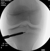
Epiphysiodesis with percutaneous drilling
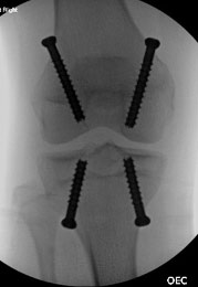
Epiphysiodesis with PETS (Percutaneous Transphyseal Screws)
Hemi-Epiphysiodesis
Techniques for hemi-epiphysiodesis include 1) guided growth plates/screws and 2) percutaneous transphyseal screw (PETS). Hemi-epiphysiodesis is typically an outpatient procedure that is relatively simple, safe, and has a quick recovery time. Patients may walk immediately after surgery and generally return to normal activities within a few weeks. A second surgery is typically required to remove the hardware once normal limb alignment has been achieved.
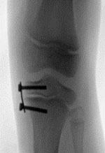
Hemiepiphysiodesis with a tension band plate
Acute Bone Shortening
Bone Lengthening
The process of gradually lengthening a bone is called distraction osteogenesis. Simply put, the surgery involves carefully cutting the bone through a small incision (<1cm) and by very slowly stretching a bone (~1mm/day), the body will create new bone in the gap formed. This has historically been accomplished using external fixators that hold the bone through pins and wires going through the skin and attaching to bars and or rings outside the limb. Newer technology evolving in the last decade now allows bone lengthening with devices that are completely on the inside of the body, making the process more tolerable for patients and with less risk of complication. With one surgery, patients can lengthen up to 8-10cm (3-4 inches). Dr. Ruggles performs bone lengthening for small leg length discrepancies as well as those with severe limb length differences that require multiple lengthenings over the course of childhood. Although bone lengthening is most often performed for patients with a leg or arm length inequality, it may also be done simultaneously in both legs for patients with conditions associated with short stature, as well as in patients that desire to be taller for cosmetic reasons.
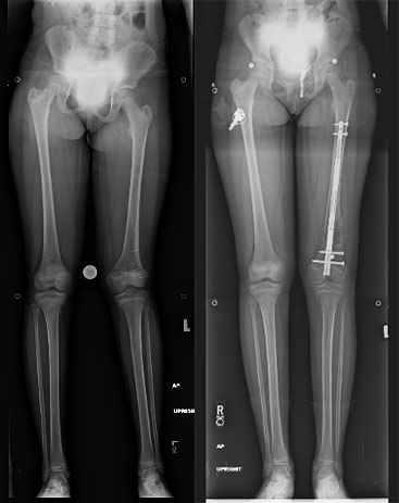
10 year old female with unequal leg lengths and deformity before and after surgery. Her leg was straightened and lengthened over 2 inches using a state-of-art internal bone lengthening device.
Deformity Correction With Hexapods
Creating Your Limb Reconstruction Plan
Contact Pediatric Orthopedics of SW FL Today
If you and your family are interested in learning more about limb lengthening and deformity correction, make an appointment with Peds Ortho and we will personally explain bone lengthening and deformity correction in detail, and create a Limb Reconstruction Plan unique for you and your child.